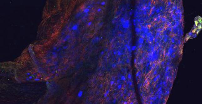
Rat lumbar dorsal root ganglia infected with dual fluorescent Varicella
Zoster Virus (red, green) and retrogradely labeled with FastBlue (blue)
resulting from footpad infection. Image from MVM Graduate Student Jean-Marc Guedon in Dr. Kinchington's lab.
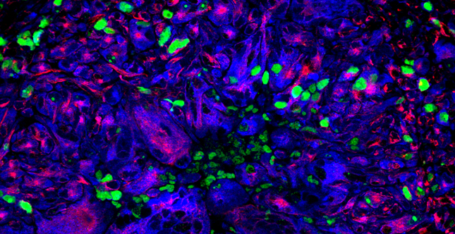
Granulomas
Pro-inflammatory and anti-inflammatory factors in granulomas - red: arginase 1, green: neutrophils, blue: CD163+ macrophages. Image from Joshua Mattila in Dr. JoAnne Flynn's laboratory.
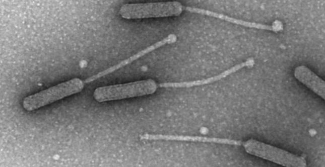
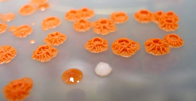
Mycobacteriophage Corndog
Mycobacteriophage Corndog. Image from Dr. Graham Hatfull's laboratory.
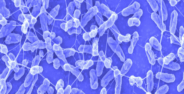
Serratia marcescens biofilm
Scanning electron micrograph of a Serratia marcescens biofilm (false colored). Image from the lab of Dr. Robert Shanks.
U2OS cells
Human osteosarcoma U2OS cells transfected with the RIG-I like receptor
(RLR) adaptor mitochondrial antiviral signaling (MAVS, in green) and
costained with Mitotracker Red. Image from Dr. Carolyn Coyne's lab.
Rough Colony Morphology
Rough colony morphology (volcano like) of an environmental Serratia marcescens isolate and mutant derivatives (red and shiny colony, white and rough colony). Image from the laboratory of Dr. Robert Shanks.
Home
***PLEASE NOTE***
THE MVM PROGRAM IS NO LONGER PART OF THE INTERDISCIPLINARY BIOMEDICAL GRADUATE PROGRAM. A NEW PROGRAM HAS REPLACED THE OLD MVM PROGRAM, THE GRADUATE PROGRAM IN MICROBIOLOGY AND IMMUNOLOGY CAN BE FOUND AT WWW.PMI.PITT.EDU